Bone Sarcoma Growth: Speed, Factors, and What It Means for You
When faced with a diagnosis like bone sarcoma, one of the most pressing, instinctive questions is often: “How fast is this growing?” It’s a natural reaction rooted in the desire to understand urgency, prognosis, and the window for intervention. Unlike a common cold or a skin rash where progression might feel more tangible, bone sarcoma growth operates on a complex biological timeline that defies simple, universal speed limits.
There’s no single answer like “it grows 2 millimeters per day.” Instead, understanding bone sarcoma growth requires navigating subtypes, tumor grades, individual biology, and the critical distinction between local expansion and metastatic spread.
Beyond a Simple Speedometer: Why “How Fast” Is Complicated
Before diving into specifics, it’s crucial to reframe the question. Bone sarcoma isn’t a single disease; it’s a family of over 50 distinct malignancies arising from bone or cartilage cells. Each subtype has inherent biological behaviors influencing its growth potential.
Furthermore, pathologists grade sarcomas based on how abnormal the cells look under a microscope (histologic grade), which is a stronger predictor of growth rate and aggressiveness than the subtype alone in many cases. A high-grade tumor, regardless of exact subtype, typically grows and spreads faster than a low-grade one.
Location also matters—a tumor in the femur (thigh bone) might present differently symptom-wise than one in a rib or the skull base due to surrounding tissue constraints and vital structures nearby. Finally, individual patient factors like age, overall health, and even specific genetic mutations within the tumor cells play a role.
Therefore, when discussing growth speed, we must consider:
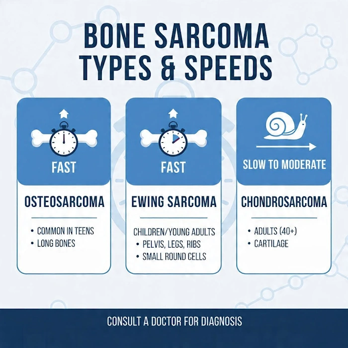
- The Specific Subtype: Osteosarcoma, Ewing sarcoma, chondrosarcoma, etc., each have typical behaviors.
- The Tumor Grade: Low-grade (slow-growing, less aggressive) vs. High-grade (fast-growing, highly aggressive).
- Local Invasion vs. Metastasis: How quickly the main tumor destroys surrounding bone/soft tissue vs. how quickly cancer cells spread to distant sites (like lungs or other bones).
- Individual Variability: No two tumors, even of the same subtype and grade, behave identically.
Let’s break down the growth patterns of the most common primary bone sarcomas, keeping these nuances firmly in mind.
Osteosarcoma: The Aggressive Adolescent (and Young Adult) Adversary
- Typical Presentation: Osteosarcoma is most common primary bone sarcoma in children, teenagers, and young adults (peak incidence 10-25 years), though a second peak occurs in older adults often associated with pre-existing bone conditions like Paget’s disease or radiation fibrosis. Favors the metaphyseal (growing end) regions of long bones around the knee (distal femur, proximal tibia) and shoulder (proximal humerus).
- Growth Characteristics: Osteosarcoma is universally considered a high-grade malignancy in its conventional form. This means the cancer cells are highly abnormal, divide rapidly, and have a significant propensity for both local destruction and early metastasis.
- Local Growth: The tumor aggressively infiltrates and destroys the bone marrow cavity (medulla) and breaks through the bone cortex (outer layer), often forming a large soft tissue mass. This process can be remarkably swift in symptomatic cases. Patients often report worsening pain over weeks to a few months before seeking help, correlating with rapid tumor expansion causing bone destruction, inflammation, and stretching of the periosteum (the bone’s sensitive outer layer). The classic “sunburst” or “Codman’s triangle” appearance on X-ray reflects rapid, disorganized new bone formation in response to the tumor’s aggressive growth – a sign of the tumor’s speed, not the tumor itself growing in that pattern. The tumor itself is laying down disorganized, malignant bone-like tissue (osteoid) at a fast pace.
- Metastatic Spread: This is where osteosarcoma’s speed is most feared. Micrometastases (tiny, undetectable clusters of cancer cells) are often present in the lungs or other bones at the time of initial diagnosis, even if standard imaging (like X-ray or CT) doesn’t show them yet. The rate at which these micrometastases grow into detectable lesions is variable but underscores the tumor’s systemic aggressiveness. Without prompt systemic treatment (chemotherapy), detectable lung metastases frequently develop within months. The median time to metastasis development in untreated high-grade osteosarcoma is often cited in the 3-6 month range, but this is highly variable and depends on tumor burden and biology.
- Grade Nuance: While conventional osteosarcoma is high-grade, rare variants exist:
- Low-grade (parosteal) osteosarcoma: Grows very slowly on the bone surface. Symptoms (pain, swelling) may develop over months to years. Local destruction is slow; metastasis is exceedingly rare if completely excised.
- Intermediate-grade (periosteal) osteosarcoma: Falls between low and high. Growth is slower than conventional but faster than parosteal. Symptoms often present over several months. Metastatic potential is lower than conventional but not zero.
- High-grade surface osteosarcoma: Similar aggressiveness to conventional osteosarcoma.
Ewing Sarcoma: The Small Cell Sprinter
- Typical Presentation: Second most common primary bone sarcoma in children and adolescents (peak 5-20 years), but also seen in young adults. Arises from primitive nerve tissue in the bone marrow. Favors the diaphysis (shaft) of long bones (femur, tibia, humerus) and flat bones like the pelvis, ribs, and shoulder blades.
- Growth Characteristics: Ewing sarcoma is defined as a small round blue cell tumor and is uniformly classified as high-grade. Its cells are primitive, divide very rapidly, and show high metabolic activity (which is why it often lights up intensely on PET scans).
- Local Growth: Like osteosarcoma, Ewing sarcoma destroys bone from within, often causing significant pain and swelling. Symptoms frequently develop over a relatively short period – often weeks to a couple of months – before diagnosis. The tumor can cause a palpable mass and may lead to pathologic fractures (breaks caused by the tumor weakening the bone) relatively quickly due to its lytic (bone-eating) nature. The speed of cortical destruction and soft tissue extension can be striking.
- Metastatic Spread: Ewing sarcoma has a high propensity for early metastasis, primarily to the lungs, other bones, and bone marrow. Micrometastases are commonly present at diagnosis. The speed of metastatic progression is a major concern. Without aggressive, upfront multimodal therapy (chemotherapy, surgery/radiation, more chemo), detectable metastases frequently appear within weeks to a few months. The tumor’s biological aggressiveness means that the window for effective local control before systemic spread closes rapidly. Studies analyzing time from symptom onset to metastasis highlight that delays in diagnosis and treatment initiation of even 6-8 weeks can significantly impact outcomes in high-risk cases, underscoring the perceived speed of threat.
- Grade Nuance: Unlike osteosarcoma, there are no recognized low-grade variants of conventional Ewing sarcoma. It is inherently high-grade. However, related tumors like Ewing-like sarcomas (with different genetic fusions) or mesenchymal chondrosarcoma may have slightly different behaviors, but Ewing sarcoma itself is uniformly aggressive.
Chondrosarcoma: The Variable Pace Cartilage Tumor
- Typical Presentation: Most common primary bone sarcoma in adults over 40 (though can occur younger). Arises from cartilage cells. Common sites include the pelvis, femur, shoulder, and ribs. Unlike osteosarcoma and Ewing’s, it rarely occurs in the metaphysis of long bones in children.
- Growth Characteristics: This is where the concept of grade becomes absolutely critical for understanding speed. Chondrosarcoma spans a wide spectrum of aggressiveness, directly correlated with histologic grade.
- Grade 1 (Low-Grade): The most common type. Cells look relatively close to normal cartilage, divide slowly. Growth is characteristically slow and indolent. Symptoms (often a dull ache or pain, sometimes a palpable mass) may be present for years before diagnosis – frequently 2-5 years or even longer. Local destruction of bone is gradual. The tumor tends to expand slowly within the bone marrow cavity, causing bone thinning and expansion rather than rapid, aggressive cortical destruction. Metastasis is very rare for pure, well-defined Grade 1 chondrosarcoma, especially if completely excised with wide margins.
- Grade 2 (Intermediate-Grade): Cells show more abnormality and increased mitotic activity (cell division). Growth is moderately paced – faster than Grade 1 but still generally slower than high-grade sarcomas like osteosarcoma or Ewing’s. Symptom duration might range from several months to a couple of years before diagnosis. Local destruction is more noticeable than Grade 1, with a higher chance of cortical breakthrough and soft tissue extension. The risk of metastasis (usually to lungs) is present but still relatively low compared to high-grade sarcomas; it might take years for metastases to become detectable after the primary tumor is established, though it can happen sooner in some cases.
- Grade 3 (High-Grade): Cells are markedly abnormal, show high mitotic activity, and may have areas of necrosis (cell death). Growth is significantly more rapid, approaching the pace seen in other high-grade sarcomas. Symptom duration before diagnosis is often shorter – weeks to several months, similar to osteosarcoma or Ewing’s in aggressive presentations. Local destruction can be fast and aggressive, with a high likelihood of cortical breakthrough and large soft tissue masses. The risk of metastasis (lungs, other bones) is substantial and can occur relatively quickly – detectable metastases may appear within months to a year or two following diagnosis if not treated aggressively, mirroring the behavior of other high-grade malignancies.
- Dedifferentiated Chondrosarcoma: A particularly aggressive variant where a low-grade chondrosarcoma component is juxtaposed with a high-grade non-cartilaginous sarcoma (often resembling osteosarcoma or undifferentiated pleomorphic sarcoma). The dedifferentiated component drives the aggressive behavior – local growth and metastasis can be very rapid (weeks to months for significant progression), carrying a poor prognosis similar to high-grade osteosarcoma.
- Mesenchymal Chondrosarcoma: A rare, highly aggressive variant (often considered high-grade) that can occur in younger patients. It tends to grow and spread rapidly, with a clinical course more akin to Ewing sarcoma or high-grade osteosarcoma.
- Low-grade (Grade 1) is notoriously slow (years for symptoms), intermediate-grade.
- (Grade 2) is moderate (months to years), and high-grade.
- (Grade 3) or dedifferentiated/mesenchymal variants are fast (weeks to months for significant progression). Assuming all chondrosarcomas grow at the same rate is a significant clinical error; grade is paramount.
Less Common Types: A Spectrum of Pace
- Fibrosarcoma of Bone (now often reclassified): Historically, primary bone fibrosarcoma was considered aggressive (high-grade like), with rapid growth (weeks-months symptoms progression). Much of it’s been reclassified with better molecular testing, but true rare primary fibrosarcomas are high-grade and fast-growing.
- Giant Cell Tumor of Bone (GCTB): Technically often considered locally aggressive but benign (though can metastasize rarely and malignant transformation occurs). Growth is variable but often moderately paced – symptoms over months to a year or two, causing bone expansion and destruction, but usually slower than high-grade sarcomas. Malignant transformation leads to high-grade sarcoma-like growth.
- Chordoma: Arises from notochord remnants, typically in the skull base or spine. Notorious for being locally aggressive but often slow-growing. Symptoms (pain, neurological deficits) can develop slowly over months to years. Local invasion is relentless but often gradual; metastasis (to lungs, liver, etc.) is uncommon but can occur, often years after the primary tumor is controlled. It’s more like a persistent weed with deep roots than a fast-spreading vine in many cases, though skull base chordomas can cause rapid neurological decline due to location.
- Undifferentiated Pleomorphic Sarcoma (UPS) of Bone / Malignant Fibrous Histiocytoma (MFH): When it arises primarily in bone (less common than in soft tissue), it is high-grade. Growth is typically rapid – symptoms over weeks to months, aggressive local destruction, and significant metastatic potential (lungs) with a timeline similar to osteosarcoma/Ewing’s (months to detect metastases without treatment).
How Doctors Actually Assess and Monitor Growth Speed (It’s Not a Ruler)
You won’t find an oncologist measuring your tumor with calipers every week to report “it grew 3mm today.” Growth assessment is indirect but sophisticated, relying on imaging and clinical correlation:
- Serial Imaging (The Cornerstone):
- X-ray: Good for showing bone destruction, new bone formation (like sunburst), cortical breakthrough, and soft tissue mass size changes over time. Comparing X-rays taken weeks or months apart is fundamental.
- MRI (Magnetic Resonance Imaging): The gold standard for assessing the extent of tumor within the bone marrow, across joints, and into soft tissues. It shows the tumor’s boundaries (often poorly defined for aggressive sarcomas) and surrounding edema (inflammation). Comparing MRI scans (e.g., pre-treatment, post-neoadjuvant chemo, surveillance) allows radiologists and oncologists to quantify changes in tumor volume or signal intensity – a direct measure of local growth or regression.
- CT Scan (Computed Tomography): Excellent for detailed bone cortex assessment (lysis, sclerosis) and, critically, for screening the lungs for metastases. Serial chest CTs are the primary way to detect and measure the growth of metastatic nodules. A nodule growing from 5mm to 8mm over 3 months indicates active metastatic growth.
- PET/CT (Positron Emission Tomography/CT): Measures metabolic activity (glucose uptake). High uptake correlates with tumor viability and aggressiveness. A decrease in SUV (Standardized Uptake Value) after chemo suggests tumor cell kill (response), while an increase or persistent high uptake suggests residual active tumor – a proxy for growth potential. It’s superb for detecting occult metastases and assessing treatment response.
- Clinical Symptoms: New or worsening pain, especially at night or not relieved by rest, increasing swelling, a new palpable mass, or onset of a pathologic fracture are all clinical indicators suggesting active local tumor growth and destruction.
- Biomarkers (Less Direct, Emerging): While no single blood test reliably tracks all bone sarcoma growth like PSA for prostate cancer, researchers investigate markers like circulating tumor DNA (ctDNA), specific enzymes (e.g., LDH, which can be elevated in high tumor burden), or cytokines. These are not yet standard for routine growth monitoring but hold promise for the future, particularly in tracking metastatic burden or treatment response.
Why Growth Rate Matters Profoundly for Treatment and Prognosis
Understanding the potential speed of growth isn’t just academic; it directly shapes critical medical decisions:
- Urgency of Intervention: For high-grade sarcomas (conventional osteosarcoma, Ewing’s, high-grade chondrosarcoma, dedifferentiated types), the rapid growth potential and high metastatic risk necessitate immediate initiation of multidisciplinary treatment. Delaying surgery to get a second opinion is reasonable, but delaying the start of neoadjuvant (pre-surgery) chemotherapy by weeks can allow microscopic metastases to proliferate and become clinically evident, significantly worsening prognosis. The speed dictates that the treatment clock starts ticking now.
- Treatment Strategy:
- Neoadjuvant Chemotherapy: Given before surgery primarily to shrink the tumor (making limb-salvage surgery more feasible) and, critically, to eradicate micrometastases early. Its efficacy is judged partly by how much tumor necrosis (dead tissue) is found in the specimen after surgery – a direct measure of how well the chemo countered the tumor’s growth potential. For slow-growing tumors (like low-grade chondrosarcoma), neoadjuvant chemo is often not used because the toxicity outweighs the minimal benefit; surgery alone is curative.
- Surgical Timing and Extent: For fast-growing tumors, surgery needs to happen promptly after neoadjuvant chemo to prevent regrowth. The surgeon aims for wide margins (a cuff of normal tissue) to ensure all microscopic extensions are removed – crucial because the tumor’s invasive front might be advancing rapidly. For slow-growing tumors, while wide margins are still ideal, there might be slightly more flexibility in planning complex reconstructions, though oncologic principles always prioritize margin status.
- Radiation Therapy: Used selectively (e.g., for Ewing’s sarcoma locally, unresectable chondrosarcoma, chordoma, or positive margins). Its role is often local control, and timing considers the tumor’s growth kinetics – needing to deliver sufficient dose to inhibit rapidly dividing cells before they repopulate.
- Surveillance Intensity: After treatment, the frequency and type of follow-up imaging (especially chest CT for lung mets) are heavily influenced by the original tumor’s grade and subtype’s known metastatic pattern and speed. High-grade osteosarcoma or Ewing’s survivors get very frequent scans (e.g., every 3 months for the first 2 years) because metastases, if they occur, tend to appear early and grow fast. Low-grade chondrosarcoma survivors might have less frequent imaging (e.g., every 6-12 months initially) because recurrences, if they happen, are often local and slow-growing, giving more time for intervention.
- Prognostic Information: Histologic grade is one of the strongest prognostic factors. High-grade tumors, by virtue of their rapid growth and metastatic potential, generally have a worse prognosis than low-grade tumors of the same anatomical site, even if the initial tumor size is similar. Knowing the grade (and thus inferred growth speed potential) helps oncologists stratify risk and tailor treatment intensity – a concept central to modern sarcoma management.
Why Early Detection Is Very Important
While we can’t change a tumor’s inherent biology, early detection dramatically impacts the effective speed at which the disease progresses to an advanced, harder-to-treat state.
- Recognizing Persistent Symptoms: Bone pain that is worse at night, not related to activity, lasts longer than a few weeks, is not relieved by rest or common painkillers (like ibuprofen/acetaminophen), or is accompanied by swelling, a lump, or unexplained weight loss/fatigue warrants prompt medical evaluation. Don’t dismiss persistent bone pain as “growing pains” (in kids/teens) or “arthritis” (in adults) without investigation, especially if it’s localized and worsening.
- Timely Medical Evaluation: Seeing a primary care doctor or orthopedic specialist promptly when symptoms persist leads to earlier imaging (X-ray, then likely MRI). The time from symptom onset to diagnosis is a critical window. For high-grade sarcomas, reducing this interval from, say, 3 months to 6 weeks can mean the difference between treating localized disease and facing detectable metastases at diagnosis.
- Avoiding Delays in Specialist Referral: Once a suspicious lesion is found on X-ray, prompt referral to a sarcoma center or orthopedic oncologist with expertise in bone tumors is vital. General orthopedists may not recognize the nuances, leading to inappropriate biopsies (which can contaminate tracts and complicate surgery) or delayed definitive care. Sarcoma multidisciplinary teams ensure the right biopsy (core needle, ideally image-guided) is done first, followed by accurate staging and treatment planning.
Living with the Uncertainty: A Nuanced Perspective
It’s natural to fixate on a specific growth rate number. However, focusing too much on a hypothetical “mm per day” can be misleading and anxiety-inducing. The reality is more nuanced and, importantly, offers grounds for hope:
- Individuality Rules: Your tumor’s behavior is unique. While statistics describe groups, your specific tumor’s genetics, microenvironment, and your body’s response play huge roles.
- Treatment Can Dramatically Alter Growth: Effective chemotherapy, targeted therapies (where available), radiation, and surgery don’t just remove existing tumor; they can halt or significantly slow the growth potential of residual cells. A tumor that would have grown rapidly without intervention can be controlled or eradicated with timely, appropriate treatment.
- Focus on Actionable Knowledge: Instead of fixating on an unknowable speed, focus on what you can control: seeking prompt evaluation for concerning symptoms, choosing an experienced sarcoma center, adhering to the recommended treatment plan (which is designed based on the tumor’s estimated aggressiveness), and maintaining open communication with your care team about symptoms and concerns.
- Advances Are Changing the Curve: Ongoing research into immunotherapy, targeted agents against specific sarcoma genetic drivers (like in certain chondrosarcomas or Ewing’s variants), and better surgical techniques continuously aims to improve outcomes, even for historically aggressive subtypes. The “speed” we associate with sarcoma today may be mitigated more effectively by treatments tomorrow.
Conclusion: Knowledge as Your Ally in Navigating Bone Sarcoma Growth
Bone sarcoma growth cannot be reduced to a single, simple number. It is a complex interplay of tumor subtype, histologic grade (the paramount indicator of aggressiveness), location, and individual biological factors.
High-grade subtypes like conventional osteosarcoma and Ewing sarcoma are characteristically fast-growing, often causing symptomatic local progression over weeks to months and posing a significant threat of early metastasis that demands urgent, intensive treatment. In contrast, low-grade chondrosarcoma exemplifies the other end of the spectrum, growing so slowly that symptoms may persist for years, often allowing curative surgery alone. Most other types fall somewhere along this spectrum, with grade being the critical discriminator.
Sources & References
- Mirabello, L., Troisi, R.J. and Savage, S.A. (2009) Osteosarcoma incidence and survival rates from 1973 to 2004: data from the Surveillance, Epidemiology, and End Results Program. Cancer, 115(7), pp. 1531–1543.
- Ottaviani, G. and Jaffe, N. (2009) The epidemiology of osteosarcoma. In: Cancer Treatment and Research. Vol. 152. Berlin: Springer, pp. 3–13.
- Bielack, S.S. et al. (2002) Prognostic factors in high-grade osteosarcoma of the extremities or trunk: an analysis of 1,702 patients treated on neoadjuvant cooperative osteosarcoma study group protocols. Journal of Clinical Oncology, 20(3), pp. 776–790.
- Godbert, Y. et al. (2016) Ewing sarcoma in adults: a retrospective study of 108 patients treated in the FIL sarcoma group. European Journal of Cancer, 57, pp. 73–81.
- Whelan, J.S. and Davis, L.E. (2014) Osteosarcoma, chondrosarcoma, and chordoma. Journal of Clinical Oncology, 32(6), pp. 587–596.
- Idowu, O.E. et al. (2012) Chondrosarcoma: a 10-year review of cases in a Nigerian teaching hospital. World Journal of Surgical Oncology, 10, p. 175.
- Fischer, S.R., Prickett, W.D. and Scarborough, M.T. (2010) Chondrosarcoma: a review of 107 cases with emphasis on prognostic factors and treatment outcomes. Journal of Surgical Oncology, 101(2), pp. 147–153.
- Picci, P. et al. (2004) Bone sarcomas in children: the Rizzoli experience. Journal of Bone and Joint Surgery (British Volume), 86(7), pp. 983–989.
- Czarnecki, J.S. et al. (2015) Molecular pathogenesis of osteosarcoma. Journal of the National Cancer Institute, 107(11), djv234.
- Suehara, Y. et al. (2015) Genetic analysis of osteosarcoma: current status and future prospects. International Journal of Clinical Oncology, 20(5), pp. 849–857.
- Gangi, A. et al. (2019) Imaging of osteosarcoma: current diagnostic approach and update. European Radiology, 29(11), pp. 6028–6040.
- Davis, A.M. et al. (2016) Radiation therapy for primary bone sarcoma: a systematic review. Radiotherapy and Oncology, 119(3), pp. 409–417.
- Tsuchiya, H. et al. (2005) Function and oncologic outcome after rotationplasty for lower-limb sarcomas. Clinical Orthopaedics and Related Research, 436, pp. 210–217.
- Lu, Y. et al. (2021) Prognostic value of histologic response in osteosarcoma: a systematic review and meta-analysis. Journal of Surgical Oncology, 123(6), pp. 1447–1458.
- Van Maldegem, A.M. and Gelderblom, H. (2018) Diagnosis and management of osteosarcoma. European Journal of Cancer Care, 27(3), e12824.