What Is Subungual Melanoma?
Subungual melanoma is a type of melanoma — the most serious form of skin cancer — that develops under or around the nail. The word ‘subungual’ simply means ‘under the nail’ in medical terminology. This cancer originates from melanocytes, which are the pigment-producing cells found in the nail matrix (the tissue at the base of the nail from which the nail grows).
Unlike other skin cancers that develop on exposed surfaces, subungual melanoma is hidden beneath the nail plate, making it harder to spot and — when missed — more likely to be diagnosed at a later stage. This is one reason why awareness of its early warning signs is genuinely important.
Subungual melanoma accounts for approximately 0.7% to 3.5% of all melanoma cases in the general population. However, it is notably more common in people with darker skin tones: it represents up to 20% of melanomas in Black Americans, 25% in Asian populations, and up to 35% in Hispanic populations, according to data referenced by the National Cancer Institute (NCI).
Types of Subungual Melanoma
Subungual melanoma falls under the broader category of acral lentiginous melanoma (ALM), a subtype of melanoma that appears on non-hair-bearing surfaces of the body — specifically the palms, soles, and nail areas. There are two primary presentations of subungual melanoma:
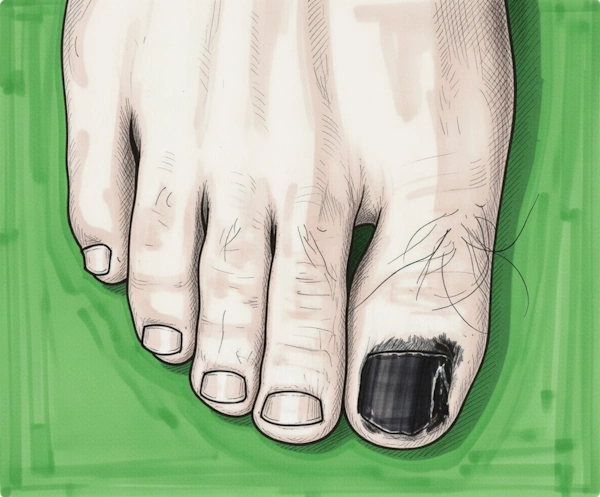
- Nail Matrix Melanoma This originates in the nail matrix — the tissue under the base of the nail — and typically presents as a dark (brown, black, or grayish) longitudinal streak running the length of the nail, called melanonychia striata or longitudinal melanonychia.
- Nail Bed Melanoma This originates in the tissue directly beneath the nail plate and may not produce pigmented streaks. It can present as a mass, lump, or ulceration under or around the nail, sometimes without any color change.
Important: The thumbnail and hallux (big toenail) are the most commonly affected nails, accounting for over 75% of subungual melanoma cases.
Subungual Melanoma Signs and Symptoms
Diagnosing subungual melanoma early can make a significant difference in outcomes. The signs are often subtle and can easily be mistaken for a bruise, fungal infection, or simply a normal nail variation.
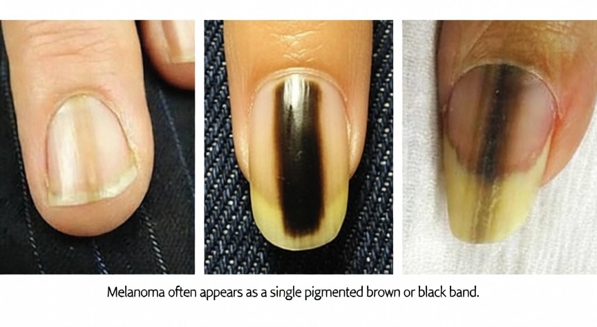
The Most Common Warning Sign: Longitudinal Melanonychia. The most recognizable early sign of subungual melanoma is a dark streak running lengthwise (from the base to the tip) along the nail.
It may be:
- Brown, black, or dark gray in color
- Single or multiple streaks
- Variable in width — often wider at the base than at the tip
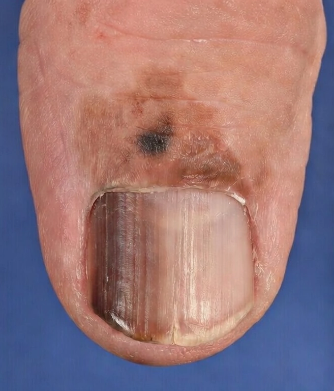
- Associated with blurring or spreading of the pigment onto the surrounding skin (this is known as the Hutchinson’s sign — see below)
The ABCDEF Rule for Nail Melanoma
Dermatologists use a modified ABCDEF rule specifically for nail changes to help identify suspicious findings. Unlike the traditional ABCDE rule for skin moles, this version is adapted for the nail:
| Letter | Stands For | What It Means |
|---|---|---|
| A | Age & African American / Asian / Hispanic origin | Most cases occur between the fifth and seventh decades of life, with a greater likelihood observed among individuals with darker skin pigmentation |
| B | Brown to Black band with Blurred borders | A pigmented streak that is brown or black, with irregular or blurry edges |
| C | Change in the nail band | The streak is growing wider, changing color, or appearing after a period of stability |
| D | Digit involved | Thumb, index finger, and big toe are highest risk; a single nail being affected increases suspicion |
| E | Extension of pigment | Pigment spreading from the nail onto the surrounding skin (Hutchinson’s sign) |
| F | Family or personal history | Personal or family history of melanoma increases risk and suspicion |
Hutchinson’s Sign
Hutchinson’s sign is a highly significant finding in subungual melanoma. It refers to the spread of brown or black pigmentation from the nail onto the adjacent skin of the finger or toe — including the nail fold (the skin at the sides and base of the nail), the cuticle, or the fingertip.
When Hutchinson’s sign is present, it strongly suggests malignancy. However, it is important to know that a pseudo-Hutchinson’s sign can occur in benign conditions as well, making clinical evaluation by a qualified professional essential.
Other Symptoms to Be Aware Of
- A nodule or raised lump under the nail
- Nail distortion, cracking, or separation from the nail bed (onycholysis)
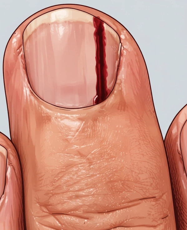
- Bleeding under the nail that does not resolve or has no clear cause
- An ulcer or sore at the fingertip or toenail that does not heal
- Destruction of the nail plate over time
- Pain or tenderness in the affected digit (this typically occurs in more advanced cases)
Key Fact: A dark streak under the nail is not always cancer. Benign causes of longitudinal melanonychia include trauma, certain medications, pregnancy, and non-cancerous nevi (moles) under the nail. Only a dermatologist can distinguish between benign and malignant causes. Do not attempt to self-diagnose.
Causes and Risk Factors
Subungual melanoma does not have a single known cause. Unlike most melanomas, ultraviolet (UV) radiation does not appear to play a significant role. Current research points to genetic mutations — particularly in genes such as KIT and NRAS — as important drivers of this cancer.
Established Risk Factors
| Risk Factor | Details |
|---|---|
| Skin color | Subungual melanoma disproportionately affects people with darker skin (Fitzpatrick skin types IV–VI). It is the most common form of melanoma in Black, Asian, and Hispanic individuals. |
| Age | Most commonly diagnosed between the ages of 40 and 70. The average age at diagnosis is around 55–65 years. |
| Personal or family history of melanoma | A prior melanoma diagnosis or a first-degree relative with melanoma increases risk. |
| Prior nail trauma | Some studies suggest repeated trauma to the nail unit may be a contributing factor, though the evidence is not conclusive. |
| Immunosuppression | Individuals with weakened immune systems (e.g., organ transplant recipients, those with HIV/AIDS) may be at higher risk. |
| Genetic mutations | Mutations in the KIT gene are found in approximately 15–20% of acral melanomas, including subungual melanoma. |
| Xeroderma pigmentosum | A rare genetic disorder that increases melanoma risk generally. |
Who Gets Subungual Melanoma?
Subungual melanoma does not discriminate by sex, and it affects both men and women at approximately equal rates. It occurs across all ethnicities, but its relative proportion of melanoma cases is significantly higher in non-white populations. This is a critical point: people with darker skin tones are often told they are not at risk for melanoma, which can delay diagnosis.
How Is Subungual Melanoma Diagnosed?
If you notice a suspicious nail change, your primary care doctor may refer you to a dermatologist — a skin specialist — or directly to a dermatologic surgeon. Here are several steps to walk through when diagnosing subungual melanoma:
Step 1: Clinical Examination
The dermatologist will examine the nail carefully, often using a dermatoscope (a handheld magnifying device with a light source, also called dermoscopy or epiluminescence microscopy). Dermoscopy allows the physician to examine the pigmentation pattern in detail without removing the nail.
Specific dermoscopic patterns associated with subungual melanoma include irregular streaks, loss of parallelism in the lines, and micro-Hutchinson’s sign (pigment on the cuticle visible only under dermoscopy).
Step 2: Biopsy
A biopsy is the only definitive way to diagnose subungual melanoma. There are several biopsy techniques used depending on the clinical presentation:
| Biopsy Type | When Used | What It Involves |
|---|---|---|
| Nail matrix biopsy | For pigmented streaks (longitudinal melanonychia) | A small portion of the nail plate is removed, and tissue from the nail matrix is sampled |
| Nail bed biopsy | For tumors on the nail bed without matrix involvement | The nail plate is elevated and a sample of the nail bed is taken |
| Excisional biopsy | For small, accessible lesions | The entire lesion is removed along with a margin of normal tissue |
| Punch biopsy | For larger lesions where full excision is not immediately possible | A circular punch tool removes a cylindrical core of tissue |
The biopsy sample is sent to a pathologist (a doctor who specializes in analyzing tissue under the microscope) for histological examination. This is what confirms whether the lesion is benign or malignant.
Step 3: Staging
If melanoma is confirmed, staging is performed to determine how far the cancer has spread. The staging system used for melanoma is the American Joint Committee on Cancer (AJCC) TNM staging system, where T = tumor thickness/characteristics, N = lymph node involvement, M = presence of distant metastases.
Melanoma Staging and Survival Rates
Stage 0~99%
Melanoma cells are confined to the outermost layers; no invasion
Stage I~90-95%
Tumor is thin (≤2 mm), localized, no lymph node involvement
Stage II~68-85%
Tumor is thicker or has ulceration, but no lymph node spread
Stage III~40-70%
Melanoma has spread to nearby lymph nodes or lymphatic channels
Stage IV~15-20%
Melanoma has spread to distant organs (liver, lungs, brain)
Note: Survival rates are general estimates based on population data. Individual outcomes depend on many factors including age, overall health, tumor genetics, and response to treatment. These figures are sourced from NCI SEER data.
Additional Diagnostic Tests
Depending on the stage and clinical picture, the following tests may be ordered:
- Sentinel lymph node biopsy (SLNB): A procedure to check if cancer has spread to the nearest (sentinel) lymph node. Often performed for tumors thicker than 0.8 mm.
- Imaging scans: CT scan, PET scan, or MRI to look for distant metastases in Stage III or IV disease.
- Genetic/molecular testing: Testing the tumor for specific mutations (e.g., BRAF, KIT, NRAS) to guide targeted therapy decisions.
- Blood tests: LDH (lactate dehydrogenase) levels can be elevated in metastatic melanoma.
Subungual Melanoma Treatment
Treatment of subungual melanoma depends on the stage at diagnosis, the location and size of the tumor, the presence of metastasis, and the individual patient’s overall health and preferences. A multidisciplinary team — typically including a dermatologist, surgical oncologist, medical oncologist, and sometimes a plastic surgeon — coordinates care.
Surgery: The Primary Treatment
Surgery is the mainstay of treatment for localized (non-metastatic) subungual melanoma. The goal is complete removal of the tumor with clear margins (no cancer cells at the edges of the removed tissue).
| Surgical Approach | When Used | Description |
|---|---|---|
| Wide local excision (WLE) | Preferred approach for thin, early-stage tumors | Removal of the tumor plus a surrounding margin of normal tissue. May preserve the digit. |
| Mohs micrographic surgery | Selected cases, nail unit preservation desired | Layer-by-layer removal of tissue with real-time microscopic examination. Tissue-sparing. |
| Amputation (digit or ray) | Historically standard; now increasingly questioned | Removal of part or all of the affected finger or toe. Evidence suggests it does not improve survival compared to WLE. |
| Nail unit excision | Early-stage lesions confined to the nail unit | Complete removal of the nail plate, matrix, and nail bed, followed by reconstruction. |
Important note on amputation: For decades, amputation was considered the standard of care for subungual melanoma. Current evidence — including data reviewed by the National Cancer Institute — suggests that for appropriately selected patients with early-stage disease, limb-sparing surgery (wide local excision) offers equivalent survival outcomes with significantly better functional and quality-of-life results. Discuss all surgical options with your surgeon.
Adjuvant Therapies (Additional Treatments After Surgery)
For higher-risk or more advanced cases, additional treatments may be recommended following surgery:
Immunotherapy
Immunotherapy uses the body’s own immune system to fight cancer. For melanoma, checkpoint inhibitors have transformed treatment outcomes over the past decade. The main agents used include:
- Pembrolizumab (Keytruda): Anti-PD-1 antibody, FDA-approved for melanoma
- Nivolumab (Opdivo): Another anti-PD-1 antibody
- Ipilimumab (Yervoy): Anti-CTLA-4 antibody, often combined with nivolumab for advanced disease
These drugs work by removing the ‘brakes’ on the immune system, allowing it to better recognize and destroy melanoma cells.
Targeted Therapy
Targeted therapy is used when the tumor has specific genetic mutations. For subungual (acral) melanoma, KIT mutations are particularly relevant:
- Imatinib (Gleevec) and nilotinib: KIT inhibitors that can be effective when a KIT mutation is present
- BRAF/MEK inhibitors (e.g., vemurafenib, dabrafenib, trametinib): Used when BRAF V600 mutations are present, which is less common in acral melanomas than in other subtypes
Radiation Therapy
Radiation is not a primary treatment for melanoma but may be used in specific situations, such as in cases with positive lymph nodes after lymph node dissection, or for palliation (symptom control) in patients with distant metastases affecting quality of life.
Clinical Trials
Given that acral melanoma (including subungual melanoma) is less common than other melanoma subtypes and has a different molecular profile, clinical trials investigating new approaches are especially valuable. Your oncologist can advise on available trials. ClinicalTrials.gov (maintained by the National Institutes of Health) is the authoritative resource for finding active trials.
Is Subungual Melanoma Curable Or Deadly?
Prognosis in subungual melanoma depends on multiple factors. The single most important prognostic factor is the Breslow thickness of the tumor — how deep the melanoma cells have penetrated into the tissue at the time of diagnosis.
| Prognostic Factor | Impact on Outcome |
|---|---|
| Breslow thickness (tumor depth) | The most critical factor. Tumors <1 mm thick have significantly better outcomes than deeper tumors. |
| Ulceration | Presence of ulceration (breakdown of the skin over the tumor) worsens prognosis and upstages the tumor in AJCC staging. |
| Mitotic rate | Higher number of dividing cells per area (mitoses/mm²) indicates more aggressive behavior. |
| Lymph node status | No lymph node involvement = significantly better prognosis. |
| Distant metastasis | Presence of metastasis to distant organs significantly worsens prognosis. |
| Stage at diagnosis | Earlier stage at detection is the strongest determinant of favorable outcomes. |
| Patient age and comorbidities | Younger, healthier patients generally tolerate treatment better and have improved outcomes. |
Common Reasons for Delayed Diagnosis
- Misdiagnosed as nail trauma or subungual hematoma (blood under the nail)
- Misdiagnosed as onychomycosis (nail fungal infection)
- Dismissed as a benign mole or normal nail variation
- Concealed by nail polish, preventing visual detection
- Limited awareness among patients and sometimes non-specialist clinicians
- Lower clinical suspicion in populations not traditionally associated with high melanoma risk
Conditions That Can Look Like Subungual Melanoma
Many benign and malignant conditions can mimic subungual melanoma. This is why professional evaluation and biopsy are essential — visual inspection alone, even by trained physicians, is not sufficient for definitive diagnosis.
| Condition | How It Can Mimic Subungual Melanoma | Key Distinguishing Features |
|---|---|---|
| Subungual hematoma | Dark streak or discoloration under the nail | Typically has a history of trauma; moves distally as the nail grows; color may be reddish-brown initially |
| Benign nail nevus (nail mole) | Longitudinal melanonychia — similar brown/black streak | Usually stable over time; homogeneous color and regular parallel lines on dermoscopy |
| Onychomycosis (nail fungus) | Nail discoloration, thickening, and destruction | Typically yellow-white-brown; no pigmented streak; confirmed by fungal culture or nail clipping microscopy |
| Subungual exostosis | Nail elevation, mass under the nail | Usually bony; X-ray shows underlying bony outgrowth; not pigmented |
| Pyogenic granuloma | Vascular, fleshy mass under or around the nail | Bright red, bleeds easily; no longitudinal melanonychia |
| Squamous cell carcinoma of the nail unit | Nail destruction, ulceration, mass | Often associated with HPV (human papillomavirus); biopsy distinguishes definitively |
| Drug-induced melanonychia | Dark nail streaks from medications | Bilateral and multiple nails affected; associated with specific drugs (hydroxyurea, chemotherapy agents, antimalarials, AZT) |
| Ethnic/physiologic melanonychia | Multiple dark streaks in people with darker skin | Multiple nails, bilateral, stable, regular lines on dermoscopy; more common in dark-skinned individuals |
After Treatment: Monitoring and Follow-Up
After treatment for subungual melanoma, ongoing surveillance is an essential part of care. The risk of recurrence and the development of a second primary melanoma are real concerns that need to be managed with regular follow-up appointments.
Recommended Follow-Up Schedule
Follow-up schedules vary by institution and by the stage of disease, but general guidelines from the National Comprehensive Cancer Network (NCCN) and other bodies suggest:
| Stage at Diagnosis | Follow-Up Frequency | Duration |
|---|---|---|
| Stage 0–I (low risk) | Every 6–12 months | 5 years; then annually as clinically indicated |
| Stage II (intermediate risk) | Every 3–6 months | First 3 years; then every 6–12 months to year 5; then annually |
| Stage III–IV (high risk) | Every 3 months | First 2–3 years; frequency reduces if no recurrence |
What Follow-Up Visits Involve
- Full skin and nail examination by a dermatologist
- Lymph node palpation (physical examination of lymph nodes)
- Imaging studies (CT, PET, or MRI) for Stage III–IV patients or if symptoms arise
- Blood tests (LDH, full blood count) for monitoring in advanced disease
- Discussion of symptoms, side effects from ongoing treatments, and psychological wellbeing
Note: After treatment, make it a habit to perform monthly self-examinations of all your nails. If you notice any new streaks, color changes, or nail abnormalities, contact your dermatologist promptly rather than waiting for your next scheduled appointment.
Prevention and Early Detection
Because subungual melanoma is not strongly linked to UV exposure, traditional melanoma prevention strategies (such as sunscreen use) are not specifically protective against it. However, early detection remains the most effective strategy for improving outcomes.
How to Examine Your Nails
You can perform a simple nail self-examination at home. This takes only a few minutes and should be done monthly:
- Remove all nail polish from fingernails and toenails before examining
- In good lighting, examine all 20 nails — including the underside of the nail and the cuticle
- Look for any new dark streaks, especially brown or black longitudinal bands
- Note any streak that is widening, changing color, or developing irregular borders
- Check the skin immediately around each nail for any pigment spread onto the surrounding skin
- Note any nail that is lifting, thickening, or showing unexplained bleeding
Who Should Be Particularly Vigilant?
- Individuals with darker skin tones (Black, Asian, Hispanic, and other non-white ethnicities)
- If you had it personally or have family cases of malignant melanoma
- Individuals over age 40, particularly those who have noticed any nail changes
- People who are immunocompromised
- Anyone who has noticed a dark streak under the nail that has been present for more than a few weeks without a clear history of trauma
What To Do If You Were Diagnosed With Subungual Melanoma
A melanoma diagnosis affects more than just the body. Anxiety, fear of recurrence, and concerns about treatment side effects and functional outcomes (especially regarding potential surgery to the hand or foot) are all normal and valid responses.
It is completely normal to feel emotional after a diagnosis. Some people find that learning about their condition in detail — as you are doing right now — helps them feel more in control. Others prefer to focus on next steps without taking in all the information at once. Both approaches are valid.
Consider asking your healthcare team:
- What stage is my melanoma, and what does that mean for my prognosis?
- What are all my treatment options, including the most conservative approaches?
- What is the likelihood of recurrence, and how will we monitor for it?
- Are there clinical trials I might be eligible for?
- Can you refer me to a mental health professional or support group that specializes in cancer care?
Functional Concerns After Surgery
If surgery is required, especially on the thumb or big toe, you may have concerns about function and appearance. Modern surgical approaches — including nail unit sparing procedures and reconstructive options — have significantly improved both cosmetic and functional outcomes compared to historical approaches. Discuss reconstructive options with your surgeon before the procedure.
Support Resources
- American Cancer Society (cancer.org): Support programs, financial assistance resources, and patient navigation
- Melanoma Research Foundation (melanoma.org): Patient resources, clinical trial finder, and support communities
- National Cancer Institute (cancer.gov): Evidence-based information on melanoma treatment, clinical trials, and support services
- CancerCare (cancercare.org): Free professional counseling, support groups, and financial assistance programs
Frequently Asked Questions
Can subungual melanoma be benign?
No. Subungual melanoma is by definition malignant — it is a cancer. The word “melanoma” itself refers to a malignant tumor of melanocytes. There is no such thing as a benign subungual melanoma.
What people sometimes confuse this with is benign longitudinal melanonychia — a dark streak under the nail that looks similar but is not cancer. This can be caused by a benign nail nevus (a mole in the nail matrix), trauma, certain medications, pregnancy, or simply normal pigmentation in people with darker skin. These benign conditions do not become subungual melanoma, though a nail nevus, like any nevus elsewhere on the body, carries a very small risk of malignant transformation over time.
There is also a pre-invasive stage called melanoma in situ (Stage 0), where the malignant cells are present but have not yet invaded deeper tissue. This is technically still malignant — it is cancer — but it is confined to the surface layer and has not spread, making it highly treatable with excellent outcomes.
How to recognize early subungual melanoma?
Early subungual melanoma most commonly appears as a single dark streak running lengthwise along the nail — from the base to the tip. This is called longitudinal melanonychia. In the early stages, it may look subtle and unremarkable, which is why it is so often missed.
Specific signs that distinguish an early melanoma streak from a benign one include:
- The streak is brown or black rather than pale tan or gray
- It appears on a single nail rather than multiple nails
- The borders are irregular, blurred, or uneven rather than clean and parallel
- The streak is wider at the base (near the cuticle) than at the tip — this triangular or wedge shape is a red flag
- The color within the streak is uneven or variegated rather than uniform throughout
- The streak is new in an adult with no history of trauma or medication that could explain it
- Faint pigment appears on the cuticle or the skin beside the nail — this early form of Hutchinson’s sign can sometimes only be seen under dermoscopy (called micro-Hutchinson’s sign) and is one of the most important early indicators a dermatologist looks for
Very early subungual melanoma confined to the nail matrix may produce only a faint, narrow streak. This is why any new unexplained dark streak in an adult — even a thin one — warrants professional evaluation rather than watchful waiting at home.
Does subungual melanoma hurt?
In most cases, early subungual melanoma does not cause pain. This is one of the most dangerous aspects of the condition — because it is painless in the beginning, people rarely feel urgency to seek medical attention.
Pain, tenderness, or soreness in the affected digit typically indicates more advanced disease. As the tumor grows deeper into the nail bed tissue or begins to destroy the nail structure, pressure and discomfort can develop. Pain may also arise when the nail plate begins to separate from the nail bed (onycholysis), or when an ulceration forms under or around the nail.
So the absence of pain is not reassurance that a suspicious nail change is harmless. Most people who are eventually diagnosed with early subungual melanoma report no discomfort at all — they noticed only a visual change in the nail.
How fast does subungual melanoma grow?
Subungual melanoma is generally considered a slow-growing cancer, but this varies considerably between individuals and tumor subtypes, and “slow” does not mean safe when left unaddressed.
The acral lentiginous subtype — which includes subungual melanoma — is known for a prolonged radial (horizontal) growth phase before it transitions to a vertical growth phase in which the tumor penetrates deeper into tissue and carries higher metastatic risk. This early horizontal phase can last months to years, which is part of why some patients live with an undiagnosed streak for a long time before it is recognized as malignant.
However, the timeline is not predictable or consistent:
- Some tumors remain in the superficial radial growth phase for several years
- Others progress to deeper invasion within months
- Once vertical growth begins — meaning the tumor is growing down into the nail bed and surrounding tissue rather than spreading along the surface — the risk of lymph node involvement and metastasis increases substantially
- The transition from in situ (Stage 0) to invasive melanoma can happen without any obvious outward change in appearance
What is the beginning of subungual melanoma?
Subungual melanoma begins at the cellular level, in the nail matrix — the tissue beneath the base of the nail where new nail cells are produced. Melanocytes (the pigment-producing cells present in the nail matrix) undergo a malignant transformation, most often driven by mutations in genes such as KIT or NRAS.
In its very earliest form — melanoma in situ — the abnormal melanocytes are present and multiplying but have not yet broken through the basement membrane into deeper tissue. At this stage, the tumor is entirely contained.
Visibly, the very first sign is usually a faint, narrow longitudinal streak — sometimes so subtle it can be overlooked or dismissed as a normal variation. The streak forms because the abnormal melanocytes in the nail matrix produce excess pigment that becomes incorporated into the growing nail plate, leaving a colored track as the nail grows outward.
Over time, as the tumor progresses:
- The streak typically widens and may develop irregular or blurry borders
- The color may become darker or more uneven
- The pigment may begin to spread onto the surrounding skin (Hutchinson’s sign), indicating the tumor has extended beyond the nail matrix
- The nail itself may begin to thicken, distort, or lift from the nail bed
- In later stages, a palpable mass or ulceration may form beneath or around the nail
Is a dark streak under my nail always cancer?
No. The majority of dark streaks under nails are benign. In people with darker skin tones, multiple dark nail streaks are particularly common and are usually normal (physiologic melanonychia). However, any new, single, widening, or irregularly pigmented streak — particularly in an adult — should be evaluated by a dermatologist. Only a professional examination and, if needed, a biopsy can determine whether a streak is benign or malignant.
My dark nail streak appeared after I bumped my finger. Should I still see a doctor?
If you experienced a clear, significant trauma (like dropping something heavy on your finger) and a dark streak appeared soon after, it is likely a subungual hematoma (bruise). A hematoma typically moves toward the tip of the nail as the nail grows out and fades over a few weeks. If the streak does not move with nail growth, does not fade, or continues to spread, see a dermatologist. When in doubt, it is always safer to have it checked.
Can nail polish cause or hide subungual melanoma?
Nail polish does not cause subungual melanoma. However, wearing opaque nail polish continuously without periodic inspection of your nails can delay detection of an early streak. It is a good practice to remove nail polish periodically and inspect your nails — perhaps once per month.
I have dark skin. Am I at higher risk?
People with darker skin tones are at lower overall risk for melanoma than those with lighter skin. However, when melanoma does occur in people with darker skin, it is disproportionately likely to be the acral lentiginous type — which includes subungual melanoma. Additionally, delayed diagnosis is significantly more common in non-white patients, partly due to lower awareness and lower clinical suspicion. This makes nail self-examination and appropriate vigilance particularly important for these populations.
Does subungual melanoma require amputation?
Not necessarily. While amputation of part or all of the affected digit was historically the standard treatment, current evidence shows that wide local excision (which preserves the digit) provides equivalent survival outcomes in appropriately selected early-stage cases. The appropriate surgical approach should be discussed with an experienced surgical oncologist who specializes in melanoma.
Can subungual melanoma come back after treatment?
Yes. Like all melanomas, subungual melanoma can recur at the original site (local recurrence), in nearby lymph nodes (regional recurrence), or in distant organs (distant recurrence). This is why lifelong follow-up with a dermatologist is recommended after treatment. Early detection of recurrence allows for prompt intervention.
Is subungual melanoma hereditary?
Most cases are sporadic (not inherited). However, individuals with a personal or family history of melanoma do have an elevated risk for all types of melanoma, including subungual melanoma. If you have a family history, inform your dermatologist, who may recommend more frequent surveillance.
Quick Reference Summary
| Topic | Key Points |
|---|---|
| What it is | Melanoma (most serious skin cancer) arising under or around the fingernail or toenail |
| Who it affects | All ethnicities; proportionally more common in Black, Asian, and Hispanic individuals; affects men and women equally; most common in ages 40–70 |
| Cause | Not strongly linked to UV exposure; likely involves genetic mutations (KIT, NRAS); exact cause not fully understood |
| Most common sign | A dark (brown/black) longitudinal streak under the nail; Hutchinson’s sign (pigment spreading onto surrounding skin) |
| Diagnosis | Clinical examination + dermoscopy + nail biopsy (the only definitive method) |
| Primary treatment | Surgical excision; digit-sparing surgery increasingly preferred over amputation when feasible |
| Additional treatments | Immunotherapy, targeted therapy (for KIT/BRAF mutations), radiation for specific indications |
| Prognosis | Strongly dependent on stage at diagnosis; early-stage disease has excellent outcomes |
| Key action | Examine nails monthly; remove polish periodically; see a dermatologist promptly for any suspicious changes |
Sources & References
- American Cancer Society (2024) Subungual (nail) melanoma. Available at: https://www.cancer.org/cancer/melanoma-skin-cancer/about/what-is-melanoma.html (Accessed: 6 March 2026).
- Bradford, P.T. et al. (2009) ‘Incidence and mortality of acral lentiginous melanoma, 1986–2005, USA’, Archives of Dermatology, 145(4), pp. 427–434. DOI: 10.1001/archdermatol.2008.609. Available at: https://jamanetwork.com/journals/jamadermatology
- Fukuoka, M., Kageshita, T. and Ono, T. (2020) ‘Diagnosis and management of nail apparatus melanoma’, Cancers, 12(11), 3237. DOI: 10.3390/cancers12113237. Available at: https://www.mdpi.com/2072-6694/12/11/3237
- Haugh, A.M. et al. (2018) ‘Molecular drivers and targeted therapy in acral and mucosal melanoma’, Frontiers in Oncology, 8, 189. DOI: 10.3389/fonc.2018.00189. Available at: https://www.frontiersin.org/articles/10.3389/fonc.2018.00189
- National Cancer Institute (2025) Acral lentiginous and subungual melanoma (PDQ®) – Patient version. Available at: https://www.cancer.gov/types/skin/patient/melanoma-treatment-pdq (Accessed: 6 March 2026).
- National Comprehensive Cancer Network (NCCN) (2025) NCCN Clinical Practice Guidelines in Oncology: Cutaneous Melanoma. Version 3.2025. Available at: https://www.nccn.org/professionals/physician_gls/pdf/cutaneous.pdf
- Reilly, D.J. et al. (2021) ‘Subungual melanoma: A review of current management and outcomes’, Journal of the American Academy of Dermatology, 85(4), pp. 995–1006. DOI: 10.1016/j.jaad.2020.08.104. Available at: https://www.jaad.org
- Ruggeri, R.M. et al. (2018) ‘Clinical, dermoscopic and histopathologic features of subungual melanoma: A systematic review’, Dermatology, 234(3–4), pp. 137–144. DOI: 10.1159/000486276. Available at: https://www.karger.com/Journal/Dermatology
- Tosti, A. and Piraccini, B.M. (2017) Nail disorders: A practical guide to diagnosis and management. 4th ed. London: CRC Press.
- World Health Organization (2024) Cancer: Skin melanoma fact sheet. Available at: https://www.who.int/news-room/fact-sheets/detail/cancer-melanoma